Clinical Value of Electroencephalography in Acute Traumatic Brain Injury
Traumatic brain injury (TBI) presents a critical challenge in acute neurological care, often dubbed a “silent epidemic” due to its prevalence and complex, sometimes hidden, effects on the brain. For patients who are unresponsive or minimally conscious, clinical examinations alone may not fully capture the subtle yet crucial changes in brain function. This is where electroencephalography (EEG) monitoring offers a powerful and objective lens into cerebral activity.
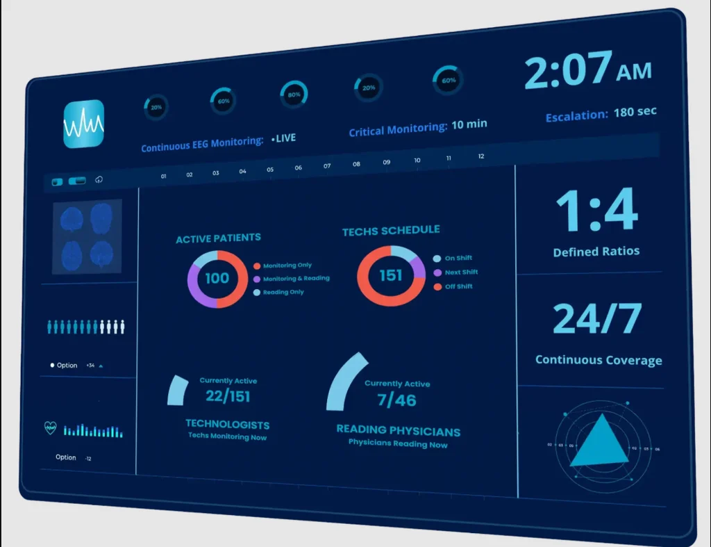
We will explore the profound role of EEG in understanding TBI. We will examine the characteristic EEG findings across different injury severities and phases, and how these patterns evolve over time. We will discover how EEG can unveil hidden consciousness in unresponsive patients, providing critical clues for prognosis and treatment. Furthermore, we will discuss the diagnostic accuracy of EEG in predicting long-term outcomes, the potential of quantitative EEG metrics as reliable biomarkers, and the practical challenges of implementing this vital monitoring in the ICU. The demand for continuous, specialized monitoring has also led to the development of solutions such as CortiCare’s tele-EEG monitoring, which help facilitate comprehensive brain surveillance.
Electroencephalography has long been recognized as a fundamental tool in neurodiagnostics, offering unparalleled millisecond-level temporal resolution to capture the dynamic electrical activity of the brain. In the context of acute traumatic brain injury, EEG provides invaluable insights into the extent of cerebral dysfunction, often revealing abnormalities that might be missed by clinical neurological examination alone. For instance, research indicates that while 86% of patients with mild TBI (mTBI) showing an abnormal neurological examination also had an abnormal EEG, a significant 23% of abnormal EEGs were present without a corresponding abnormal neurological examination. This highlights EEG’s sensitivity in detecting subtle electrophysiological changes indicative of injury.
The characteristic EEG findings in TBI vary significantly with the injury’s severity and phase, evolving dynamically over time. Immediately following a TBI, common abnormalities include a slowing of the posterior dominant rhythm (PDR), an increase in diffuse theta activity, and in more severe cases, increased delta power. These patterns reflect general cerebral dysfunction, with slower frequencies often correlating with greater injury severity. For patients experiencing unconsciousness for more than two minutes, EEG abnormalities are notably more prevalent (56%) compared to those with briefer periods (17%). Over time, these acute abnormalities tend to resolve, with the majority (90%) normalizing within one year post-injury, and a substantial portion resolving within three months.
A critical application of EEG in acute TBI is the detection of non-convulsive seizures and status epilepticus, which can significantly worsen outcomes if left untreated. Given the resource-intensive nature of continuous EEG (cEEG) monitoring, a comparison between routine and continuous approaches is often considered.
Feature Routine EEG (rEEG) Continuous EEG (cEEG) Duration Typically 20-60 minutes Hours to days (e.g., 24-72+ hours) Seizure Detection Limited, may miss transient or infrequent events High yield for non-convulsive seizures and status epilepticus IIC Detection May miss evolving ictal-interictal continuum patterns More effective in identifying severe IIC patterns Background Analysis Snapshot of brain activity Captures dynamic evolution of background rhythms over time Prognostic Value Provides some prognostic indicators Enhanced prognostic accuracy, especially with background features Resource Intensity Lower, less demanding on staff and equipment Higher, requires more equipment, staff time, and interpretation Pathophysiological Patterns Across Injury Severities
The electrophysiological landscape of the brain undergoes profound changes after TBI, reflecting the underlying neuropathology. In mild TBI, EEG often reveals subtle changes such as a slowing of the posterior dominant rhythm and an increase in diffuse theta activity. As injury severity increases, these patterns become more pronounced, with a greater prevalence of delta power, indicating more severe cortical injury and widespread brain dysfunction. Alpha suppression, where the normal alpha rhythm is diminished or absent, can also be observed, particularly in areas of focal injury or diffuse cerebral insult.
The dynamic evolution of these patterns is crucial for monitoring. For instance, the persistence of slow-wave activity beyond the acute phase, or the failure of the posterior dominant rhythm to normalize, can signify ongoing cerebral pathology or a poorer recovery trajectory. In severe TBI, patterns such as burst suppression, characterized by periods of high-amplitude activity alternating with periods of electrical silence, indicate profound brain injury and are often associated with poor prognosis.
Early Detection of Non-Convulsive Seizures and Status Epilepticus
One of the most critical roles of EEG in acute TBI is the early detection of non-convulsive seizures (NCS) and non-convulsive status epilepticus (NCSE). These events, often clinically silent, can exacerbate brain injury by increasing metabolic demand and potentially leading to further neuronal damage. Studies show that electrographic seizures can occur in 18-33% of adult moderate-to-severe TBI cases. Without continuous EEG monitoring, these events are frequently missed, as clinical signs may be subtle or absent, particularly in sedated or comatose patients.
The ictal-interictal continuum (IIC), which encompasses a range of electrographic patterns from clear seizures to ambiguous discharges, further complicates diagnosis. Severe IIC patterns, while correlating with injury severity, may not always directly predict short-term functional outcomes. However, their presence necessitates careful monitoring and often intervention. Early detection and treatment of NCS and NCSE are paramount to mitigate secondary brain injury and improve patient outcomes, potentially reducing the incidence of post-traumatic epilepsy, which affects 2% to 16% of head injury patients.
Advanced Prognostication and Cognitive Monitoring in Neurocritical Care
Beyond detecting acute pathology, EEG serves as a powerful tool for prognostication and monitoring cognitive function in patients with traumatic brain injury. Its ability to provide objective physiological data complements clinical assessments and neuroimaging, offering a more comprehensive picture of brain health and recovery potential.
Detecting Hidden Consciousness and Cognitive Processing
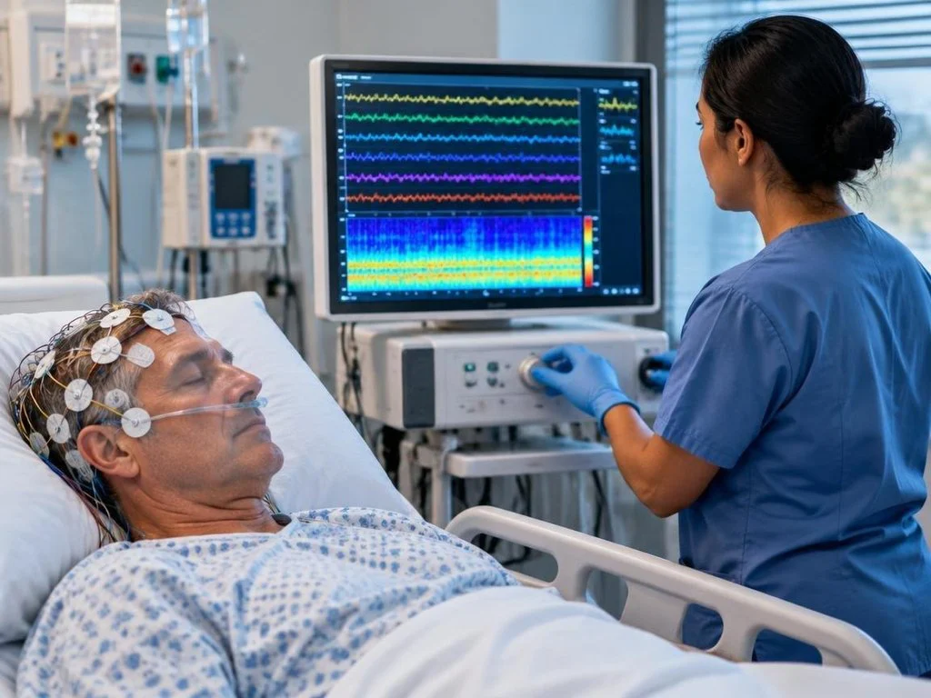
Perhaps one of the most compelling applications of EEG in TBI is its capacity to detect hidden consciousness or cognitive processing in unresponsive or minimally responsive patients. Research has shown that nearly 1 in 7 (15%) brain-injured ICU patients exhibit evidence of hidden consciousness detectable with EEG just days after injury. These patients, despite being unable to communicate or follow commands clinically, demonstrate specific EEG patterns in response to verbal commands, indicating a level of awareness previously unrecognized.
The prognostic value of these findings is substantial. Among patients with hidden consciousness signals on EEG, 50% improved and were able to follow verbal commands before hospital discharge, compared to only 26% of those without such brain activity. Even more remarkably, a year after injury, 44% of patients with early EEG evidence of hidden consciousness were able to function independently for up to eight hours each day, versus only 14% of those without such signals. This phenomenon, sometimes referred to as cognitive motor dissociation or covert consciousness, highlights the potential for EEG to redefine our understanding of consciousness and recovery trajectories in severe TBI.
Quantitative EEG Metrics and Objective Biomarkers
Quantitative EEG (qEEG) metrics offer a more objective and standardized approach to analyzing EEG data, moving beyond visual inspection to extract numerical features that can serve as reliable biomarkers. These metrics include spectral power (e.g., alpha, theta, delta bands), alpha-band variability, brain symmetry index (BSI), functional connectivity, and event-related potentials (ERPs).
For instance, a model integrating EEG features with clinical, radiological, and laboratory parameters predicted poor outcome in moderate to severe TBI with an impressive AUC of 0.89, sensitivity of 0.83, and specificity of 0.85. Even using EEG parameters alone at 72 hours post-trauma, the best prediction model achieved an AUC of 0.82, specificity of 0.83, and sensitivity of 0.74 for predicting poor clinical outcome.
Specific qEEG metrics show promise for monitoring recovery, guiding treatment decisions, and assessing response to interventions. For example, changes in ERP amplitudes and alpha-band spectral power have been identified as primary EEG outcomes in clinical trials assessing the efficacy of treatments like hyperbaric oxygen therapy in veterans with TBI. These objective biomarkers can track neurophysiological changes over time, offering a scalable alternative to subjective symptom-based assessments, especially in cases complicated by comorbid conditions like PTSD.
Implementing Continuous Protocols with CortiCare’s tele-EEG monitoring
Implementing continuous EEG (cEEG) monitoring in the intensive care unit (ICU) for TBI patients, while highly beneficial, presents a unique set of practical challenges and methodological considerations. These include managing complex ICU workflows, ensuring consistent data quality, and overcoming staffing limitations. However, advancements in remote neurotelemetry and specialized services are increasingly addressing these hurdles.
The integration of tele-EEG networks, such as those provided by CortiCare, allows for expert neurophysiologist interpretation of cEEG data from a remote location, providing critical support to bedside teams. This not only facilitates rapid triage and intervention but also ensures that specialized expertise is available 24/7, regardless of the hospital’s geographic location or internal staffing. For hospitals seeking to enhance their acute neurodiagnostic capabilities and improve patient outcomes, exploring advanced solutions like CortiCare’s tele-EEG monitoring can provide a comprehensive framework for cerebral surveillance.
Addressing Staffing Bottlenecks Using CortiCare’s tele-EEG monitoring
One of the most significant barriers to widespread cEEG implementation is the shortage of trained neurophysiologists and EEG technologists, especially for round-the-clock coverage. Tele-EEG monitoring directly addresses these staffing bottlenecks by centralizing expertise. This model enables 24/7 surveillance of multiple patients across different ICUs, providing continuous monitoring that would otherwise be impossible for many institutions.
Remote neurophysiologist access ensures that critical alerts, such as the onset of non-convulsive seizures, are promptly identified and communicated to the bedside team, allowing for timely intervention. This remote support improves interpretation turnaround times, enhances bedside efficiency by reducing the need for on-site specialists, and ultimately contributes to better patient management. The ability to deploy a robust EEG monitoring program without needing to hire additional full-time, highly specialized staff makes tele-EEG an attractive and practical solution for many healthcare systems.
Clinical Trial Integration with CortiCare’s tele-EEG monitoring
The rigorous demands of clinical trials, particularly in TBI research, necessitate highly standardized and reproducible methodologies. EEG, with its objective quantifiable metrics, is increasingly being integrated into these trials to monitor therapeutic response and track longitudinal changes. However, maintaining consistent EEG protocols across multiple sites, managing blinding, and ensuring robust artifact control can be challenging.
Tele-EEG services can play a pivotal role in clinical trial integration. By providing standardized protocols for electrode placement and data acquisition, and offering centralized, blinded interpretation by experienced neurophysiologists, tele-EEG helps ensure data integrity and consistency across multicenter trials. This is particularly valuable when assessing the efficacy of novel interventions, such as hyperbaric oxygen therapy, where subtle neurophysiological changes need to be precisely tracked. The ability to reliably monitor and analyze EEG data longitudinally, even with remote oversight, strengthens the scientific rigor and scalability of TBI clinical research.
Addressing Technical and Practical Challenges in Bedside Neurodiagnostics
While the clinical value of EEG in TBI is undeniable, its implementation in the dynamic and often chaotic environment of the ICU comes with a specific set of technical and practical challenges. These range from managing electrical artifacts to ensuring proper electrode placement and maintaining data quality over prolonged periods.
Feature Conventional Continuous EEG (cEEG) Point-of-Care / Patch-Type EEG Setup Complexity High (multiple electrodes, wires, dedicated equipment, technologist) Low (fewer electrodes, wireless, nurse-deployable) Portability Limited (requires cart-based system) High (wearable, compact, allows patient mobility) Channel Count High (16-32+ channels) Lower (e.g., 2-8 channels) Artifact Susceptibility Moderate to High (due to wires, patient movement) Moderate (can be affected by movement, but fewer wires) Data Quality Excellent (with proper setup and technologist oversight) Good (sufficient for seizure detection, background trends) Deployment Staff Trained EEG technologist ICU nurse (with minimal training) Cost Higher (equipment, technologist, neurophysiologist) Lower (equipment, less specialized staff needed for deployment) Primary Use Case Comprehensive brain monitoring, detailed analysis, prognostication Seizure detection, trend monitoring, resource-limited settings Managing Artifacts and Data Quality in the Intensive Care Unit
The ICU environment is inherently challenging for EEG recordings. Electrical artifacts from other medical equipment (e.g., ventilators, infusion pumps) can interfere with brain signals, creating noise that obscures true cerebral activity. Patient movement, tremors, and even routine nursing care can lead to electrode displacement or movement artifacts. Physiologic noise, such as muscle activity (EMG) or eye movements (EOG), also needs to be carefully managed.
To combat these issues, meticulous attention to montage standardization, proper skin preparation, and electrode application is crucial. Algorithmic filtering and advanced signal processing techniques can help mitigate some artifacts during data analysis. Regular checks of electrode impedance and careful documentation of patient activities and interventions are essential to ensure high data quality and avoid misinterpretation of baseline drift or spurious events. Blinding protocols, where interpreters are unaware of clinical details or intervention groups, also require robust artifact control to prevent bias.
Special Considerations in Veteran Populations and Comorbid Trauma
Veterans with TBI often present with unique clinical complexities, including a high prevalence of comorbid conditions such as post-traumatic stress disorder (PTSD) and chronic pain. These comorbidities can significantly impact neurobehavioral, cognitive, and functional outcomes, making TBI diagnosis and monitoring particularly challenging. For example, symptoms of PTSD can overlap with post-concussive symptoms, making it difficult to attribute specific complaints solely to brain injury.
EEG, especially qEEG, offers an objective means to assess brain function in these complex cases. While there is no definitive unique qEEG signature for mild TBI, spectral analysis and ERPs can provide insights into neurophysiological alterations that may correlate with specific symptoms or treatment responses. Research into interventions like hyperbaric oxygen therapy for blast-related TBI in veterans often uses EEG to track neurophysiological changes, providing objective measures of treatment efficacy that complement subjective symptom reporting. The ability of EEG to capture subtle changes in brain activity can aid in distinguishing the neurophysiological impact of TBI from that of comorbid conditions, guiding more personalized treatment planning and neurorehabilitation strategies.
Frequently Asked Questions About Acute EEG Monitoring
What is the primary clinical difference between continuous and routine EEG in acute TBI?
The primary clinical difference lies in their diagnostic yield and temporal resolution for detecting transient or infrequent events. Routine EEG (rEEG), typically lasting 20-60 minutes, provides a snapshot of brain activity. While useful for initial assessment and detecting gross abnormalities, it has a limited ability to capture non-convulsive seizures (NCS) or non-convulsive status epilepticus (NCSE), which can be intermittent or evolve over hours. Continuous EEG (cEEG), on the other hand, monitors brain activity over extended periods (e.g., 24-72+ hours), significantly increasing the likelihood of detecting these clinically silent but damaging events. Studies have shown that cEEG identifies additional interictal epileptiform discharges and seizures that are missed by shorter recordings, making it superior for comprehensive cerebral surveillance in high-risk TBI patients.
How does EEG detect covert consciousness in clinically unresponsive patients?
EEG detects covert consciousness through the analysis of specific brain activity patterns in response to external stimuli, particularly verbal commands. In patients who are clinically unresponsive (i.e., unable to move or communicate), researchers present motor commands (e.g., “move your right hand”) and look for corresponding, task-specific changes in EEG signals. These changes typically involve spectral modulation, such as an increase in alpha or theta power, or specific event-related potentials (ERPs) that are known to be associated with motor imagery or command comprehension. The presence of these active paradigms, even without overt behavioral response, indicates that the brain is processing and attempting to execute the command. This detection of hidden consciousness provides crucial prognostic stratification, as these patients often have a significantly higher chance of functional recovery.
Which quantitative EEG metrics provide the highest prognostic value after trauma?
Several quantitative EEG (qEEG) metrics have shown high prognostic value after traumatic brain injury, particularly when integrated into multimodal prediction models. Among the most consistently reported are alpha-band variability, relative alpha power, and absolute beta power, which contribute significantly to predicting poor clinical outcomes. Other important metrics include the brain symmetry index (BSI), which assesses hemispheric imbalances, and various measures of background reactivity and organization (e.g., presence of posterior dominant rhythm, N2 sleep transients, predominant delta activity, discontinuous background). Event-related potentials (ERPs) also serve as valuable objective biomarkers, with changes in their amplitudes and latencies correlating with injury severity and treatment response. The integration of these qEEG parameters with clinical scores (like the IMPACT score) and neuroimaging data creates robust prognostic models, enhancing the accuracy of outcome prediction for TBI patients.
Conclusion
The journey through the complexities of traumatic brain injury underscores the indispensable role of EEG monitoring in modern neurocritical care. From revealing characteristic pathophysiological patterns across varying injury severities to the critical early detection of non-convulsive seizures, EEG provides a dynamic window into the injured brain. Its advanced capabilities extend to unveiling hidden consciousness in unresponsive patients, offering profound implications for prognostication and family counseling. Furthermore, quantitative EEG metrics are emerging as powerful, objective biomarkers, guiding treatment decisions and assessing the efficacy of interventions.
While practical challenges persist, particularly in the demanding ICU environment, technological advancements in tele-EEG and portable systems are paving the way for more scalable and accessible cerebral surveillance. These innovations are crucial for optimizing neurocritical workflows, ensuring timely interventions, and ultimately improving clinical recovery trajectories for TBI patients. As we move forward into October 2026, the continued integration of EEG, supported by remote monitoring solutions, promises to refine our understanding and management of TBI, fostering secondary prevention and enhancing patient outcomes across the globe.
Read more : Essential Pet Care Tips to Keep Your Furry Friends Healthy and Happy